Optometrists, Ophthalmologists, Opticians

Keratoconus

Keratoconus is a terrifying diagnosis to those that have experienced it. To compound issues, many patients complain that they had poor initial treatment due to a lack of understanding about the disease. If proper treatment is not achieved, individuals may experience a rapid deterioration in their ability to see. This leads to a reduced quality of life. You can reduce the stress related to a keratoconus diagnosis and increase the benefits of treatment by understanding your treatment options.
 

Understanding Keratoconus

Keratoconus is an eye disease that causes the cornea to thin and bulge. This bulge generally takes on the appearance of a cone. As light enters the eye, it becomes distorted by the cone causing vision abnormalities.

Modern research is connecting keratoconus with an enzyme imbalance in the cornea. This imbalance leaves the eye susceptible to oxidative free radicals. Keratoconus has also been linked to UV damage, excessive eye rubbing, poorly fitting contacts, and chronic eye irritation.
 

Treatment Options

While your eye professional will have the best understanding of what treatment option is right for you, we have compiled ten of the most common treatments here.

  • Corneal Cross-linking (CXL) – There are two different types of this procedure, but they both introduce riboflavin to the cornea in order to strengthen the corneal tissue and stop the bulging from progressing.
  • Custom Soft Contact Lenses – Soft contacts are generally more comfortable to wear than gas permeable lenses. Recently, some contact companies have been able to create a contact specifically to correct the issues related to mild and moderate cases of keratoconus.
  • Gas Permeable Contact Lenses – Gas permeable lenses are a hard contact lens that physically forces the eye to adhere to the lens shape. This allows for the correction of keratoconus. The fit is often time-consuming and may take several different lenses to achieve the proper fit.
  • Piggybacking Contact Lenses – This method is used for individuals who require a gas permeable lens but cannot tolerate wearing rigid contacts. Piggybacking utilizes a soft lens placed on the eye first, and then a gas permeable lens is placed over the top. This offers the comfort of soft contacts with the rigidity and clarity of the gas permeable lenses.
  • Hybrid Contact Lenses – Hybrid contact lenses were designed specifically for keratoconus. This technology blends a rigid contact lens center with a softer edge, or skirt, of the contact
  • Scleral and Semi-Scleral lenses – These lenses are gas permeable lenses but cover a larger area of the eye than a standard rigid lens. These lenses don’t put pressure onto the cone shape of the eye. The reduced pressure results in a more comfortable fit for patients.
  • Prosthetic Lenses – This lens is used specifically for patients that have very advanced keratoconus and have ruled out other options. The advanced scleral lens also doubles as a protective prosthetic shell. There are special requirements to qualify for this lens though, so check with your eye care professional if this is an option for you.

​​​​​​​

  • Intacs – These implants are surgically placed in the eye. They are a small plastic insert that is inserted into the eye, just under the surface. This option is also designed for patients who could not use other contact lens types.
  • Topography-Guided Conductive Keratoplasty – This treatment option is still being researched, but it uses the energy from radio waves and small probes to map the surface of the eye. This detailed mapping allows for an appropriate treatment plan for the patient.
  • Corneal Transplant – Patients with advanced keratoconus may no longer be able to tolerate a contact lens, or the different lens types may not be correcting the issues. This surgery removes the damaged cornea and replaces it with a healthy cornea.
     

There is hope for individuals with keratoconus. Even though the disease is degenerative, many patients report restored vision and improved quality of life when the appropriate treatment plan is in place. If you have been diagnosed with keratoconus or are having problems with your vision, see your eye care professional today.

Read More
Common Ocular Diseases

Both optometrists and ophthalmologists treat many common types of ocular disease. However, for the best outcome, it’s important to see an eye doctor regularly. They can identify any issues before they become serious problems.

Fortunately, they can treat all of the diseases mentioned below, and in some cases, you can do certain things to prevent them from developing. Look at the most recent statistics, and you’ll see why good eye health care matters.

Currently, more than 4.2 million people in the U.S. alone over the age of 40 are partially blind or have poor visual acuity. Although a lot of things cause these problems, the ocular diseases listed below are the most common.

Macular Degeneration

This is commonly referred to as “age-related macular degeneration” because it affects seniors. Not only does it cause blurriness and distortion but left untreated, individuals lose their central vision. In other words, they are unable to see anything through the center portion of the eye.

Two types of this ocular disease exist. First, wet macular degeneration means that abnormal blood vessels that are located behind the retina grow under the macular. Along with leaking blood and fluid, this leads to scarring and, sometimes, permanent damage. Second, dry macular degeneration progresses slowly as part of the natural aging process. Typically, it affects both eyes at some point.

Cataracts

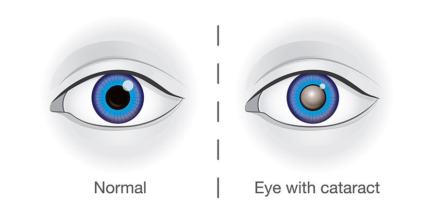
Roughly 20 million people in the U.S. over the age of 40 have cataracts in either one or both eyes. While they can develop in children, teens, and young adults, cataracts are most often associated with age. With this, a film covers the eye, which, in turn, makes everything appear blurry.

Of all the different kinds of ocular diseases that lead to blindness worldwide, cataracts rank number two. Fortunately, an eye doctor can remove the damaged lens, followed by implanting an artificial one. After recovery, patients see amazingly well.

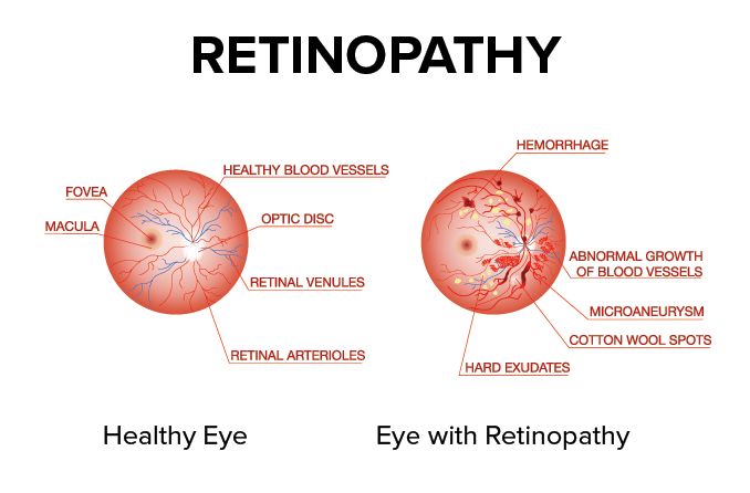
Diabetic Retinopathy

If you have diabetes, then you’re at risk of developing this ocular disease. This particular disease causes progressive damage to the retina’s blood vessels. The first stage consists of mild non-proliferative retinopathy and then moderate non-proliferative retinopathy, which blocks some of the vessels.

Then, it moves into stage three or severe non-proliferative retinopathy, which means more blood vessels become blocked. The fourth and final state, proliferative retinopathy, is the most advanced. Although Diabetic Retinopathy does affect just one eye on occasion, it typically involves both eyes.

Start by improving your overall health. Eat balanced meals, keep your blood pressure and cholesterol levels down, and take insulin. In addition, regular exercise, losing weight, and giving up smoking all make a huge difference. From there, a qualified eye doctor can provide you with treatment options to reduce the risk of losing your vision.

Glaucoma

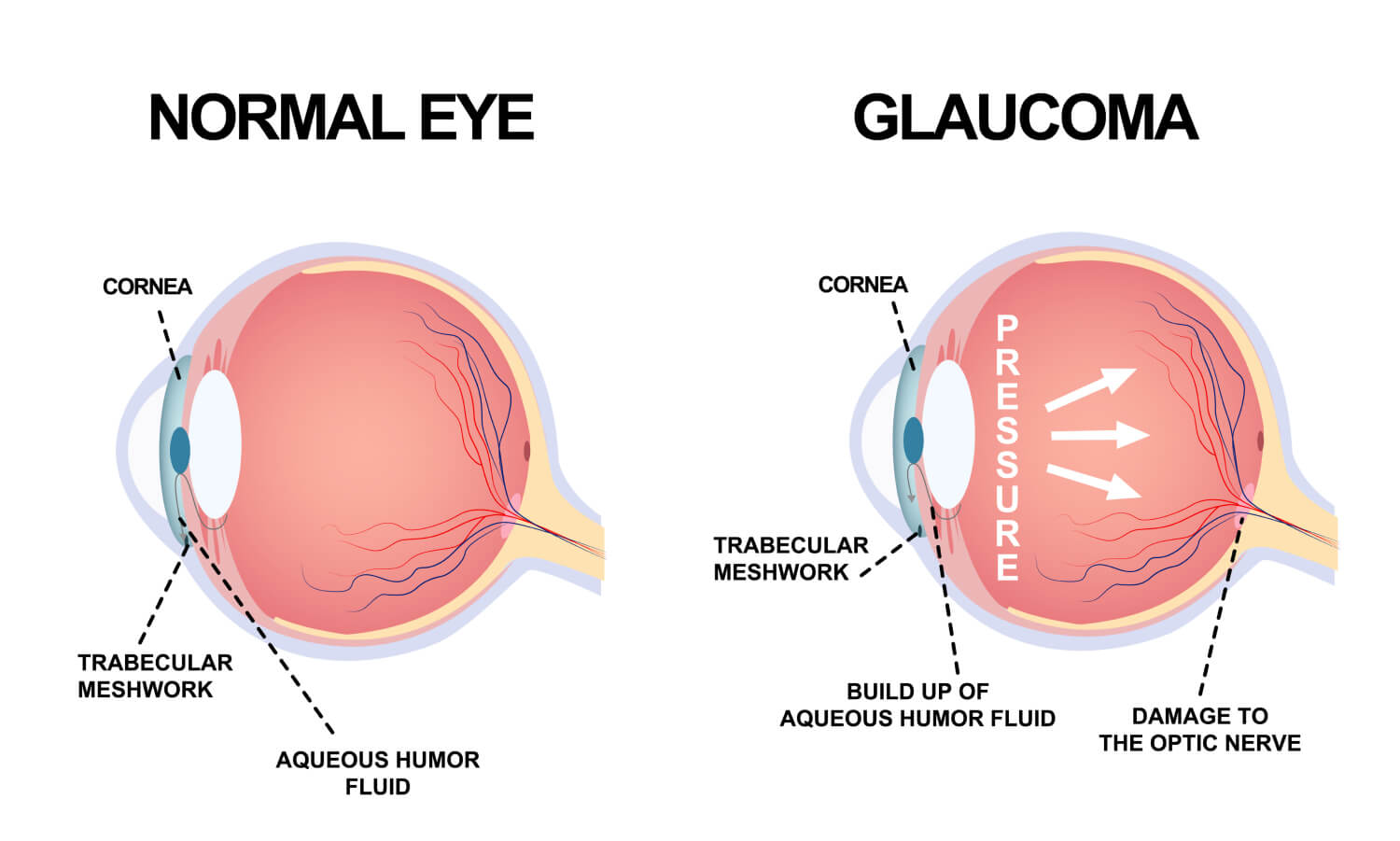
Many people think glaucoma is one type of ocular disease. However, it’s a group of diseases that cause damage to the optic nerve. When that happens, people face the risk of losing their sight completely. With glaucoma, the fluid pressure inside the eyes gradually rises.

There are also two categories of glaucoma: open-angle and closed-angle. Not only is open-angle glaucoma chronic, but it also progresses slowly. Often, a person can have this type without knowing it. Unfortunately, they don’t realize there’s an issue until they have a comprehensive eye exam performed.

As for closed-angle glaucoma, it’s usually painful and it comes on suddenly. In addition, an individual can lose their vision much faster with this kind of glaucoma compared to the open-angle kind. Because this happens fast and involves pain, it’s diagnosed much quicker as well.

For these common types of ocular diseases, it’s important to have your vision checked. If an ocular disease is diagnosed, your optometrist will be able to determine the best treatment plan for optimal eye health and vision.

Read More

Diabetic Related Eye Exams

You have almost certainly heard of diabetes, which is one of the most common chronic health conditions in the United States with an estimated 100 million adults currently living with diabetes or pre-diabetes. This metabolic disorder occurs when the body is no longer able to regulate its own blood sugar levels and requires intervention to keep them stable. Most people are aware that diabetes can have serious consequences for our health. However, you may be surprised to learn that it can also influence our vision. This is because patients who are diabetic can go on to develop a complication that is known as diabetic retinopathy. Without prompt treatment, diabetic retinopathy can cause permanent vision loss. It is for this reason that patients who suffer from diabetes are asked to attend regular diabetic-related eye exams. 
 

What Is Diabetic Retinopathy?

For us to be able to see clearly, our eyes need to be healthy and functioning perfectly. The most important component of our eyes are the retina. Found at the very back of the eye, the retina is a patch of light-sensitive cells that have the job of converting the light that passes into the eye into messages that are passed up the optic nerve and into our brain. Our brain then receives them and tells us what we can see and how clearly we can see it. 

The retina relies on a continuous supply of blood, which is delivered using a network of tiny blood vessels. Over time, having continuously high blood vessels can damage these blood vessels causing a leak of blood and other fluids onto the retina. If this happens, scarring may occur which could compromise the quality of your vision. 
 

Am I At Risk Of Diabetic Retinopathy?

Technically, anyone who suffers from diabetes, whether it be Type 1 or Type 2, could be at risk of developing diabetic retinopathy. However, the condition is more likely in certain situations. These include if:

  • your blood sugar levels are uncontrolled or poorly controlled
  • you have a long history of diabetes
  • you have high blood pressure (hypertension)
  • you suffer from high cholesterol 
  • you are pregnant

Regular diabetic-related eye exams will enable your eye doctor to monitor your condition and ensure that any signs of diabetic retinopathy are detected and acted upon immediately. 
 

What To Expect From Diabetic-Related Eye Exams?

The process of a diabetic eye exam is very simple and straightforward. In fact, in most instances, it is included within the other elements of comprehensive eye exam and you may not even realize that you have had a specific test to check for diabetes-related complications. 

Diabetic eye screening is non-invasive. You will be given eyedrops which will blur your vision. These may sting a little when they are administered, but this will pass within just a few moments. Once your vision is blurred, you will be asked to rest your head onto a device and stare down a lens. This leads to a camera which will take images of the backs of your eyes so that your eye doctor can assess the structures, which include the retina, for any abnormalities. You will see a flash when each image is taken, but at no point should you be in any pain. 

In addition to the images of the back of your eye being taken, you will also be given a visual acuity test. This is where you will be asked to read letters off a chart a short distance away, as well as reading from a card held in front of you. 

The information that your eye doctor will obtain from your examination will be able to tell them if you are experiencing any of the signs of diabetic retinopathy. If so, they will discuss the best way to get your condition under control. This could involve a combination of elements, including controlling your diabetes more effectively, taking medications or more invasive treatment to preserve your vision. Your eye doctor will give you more specific information based on your individual circumstances. 

If you have further questions about diabetic-related eye exams, please contact a knowledgeable eye care team. 

Read More

Pink Eye

When you were a kid, did you experience your eyes become reddish and all of a sudden, someone close to you was also suffering from it? Your eyes, as well as those who contracted it, got itchy and swollen, right? Then it must have been that you were suffering from pink eye.

Pink eye is well known as conjunctivitis and it is the infection or inflammation of the conjunctiva or the transparent membrane that serves as a covering for the white part of the eye called the sclera that lines the eyelid. In addition to inflammation, there is usually tearing in the eyes that emits a sticky discharge which develops into a crust while one is sleeping, making it difficult for the patient to open their eyes in the morning.

One thing about the pink eye, which could affect one or both eyes, is that it is highly contagious. While it is more common in children, adults can also be victims of this eye condition. Here are the 3 major causes of pink eye:
 

Bacteria

Streptococci and staphylococci are bacteria types that are most responsible for pink eye. However, chlamydia and gonococci can also cause pink eye. It is accompanied by serious eye pain, itching, swelling, redness, and discharge. The spread of bacterial pink eye is usually as a result of using personal items of infected parties, such as makeup or makeup tools that have been infected with bacteria or putting dirty hands in the eyes. If not treated, it can last for more than 10 days, but if treated, it should resolve in less than 3 days.
 

Allergies

Pink eye caused by allergies is followed by serious itching and tearing of the eyes. Pain is minimal, but it typically comes with quite a bit of discomfort. Most of the time, pink eye is accompanied by sneezing or coughing. Allergens that trigger pink eyes include grass, dust, pollen, mold, and ragweed. Allergy based pink eye is not usually contagious.
 

Viruses

Viruses such as the adenoviruses and herpes virus are the most common causes of pink eye. When a virus is the cause, there is usually a lot of teary discharge accompanied by nasal congestion, puffy eyelids, runny nose, and sharp pain. It is usually contracted from cough and sneeze droplets from an infected individual. It can take as long as 2 weeks to treat depending on the seriousness of the infection.

General Symptoms of Pink Eye:

  • Itchy eyes
  • Redness of the sclera
  • Pain
  • Watery discharge
  • Swollen eyelids
  • Hazy or blurry vision
  • Oversensitivity to light
     

Prevention

The best way to prevent pink eye is by practicing good hygiene which includes:

  • Avoid putting dirty hands in your eyes
  • Make it a habit to wash your hands often
  • Avoid sharing towels and other personal items
  • Do not use dirty items
  • Changing your pillow cover regularly
  • Do not leave a makeup item open for too long
  • Avoid sharing makeup items like eyeliners, mascara, etc.
     

Treatment Of Pink Eye

  • The treatment of pink eye is dependant on its underlying cause. If it is caused by a virus, you just might have to wait for the virus to run its course which could last for about four to seven days. Virus caused pink eye could be easily contracted so it is imperative to try and prevent further spreading. Viruses cannot be cured by antibiotics, but some antiviral drugs could be helpful.
  • Antibiotics are most effective against pink eye caused by bacteria as they reduce the lifespan of these bacteria and could come in the form of eye drops or pills. Based on the doctor’s prescription, an eye drop should be administered about four to six times daily. It is important you finish using your drugs even after the disappearance of symptoms.
  • To deal with pink eyes caused by allergies, the allergy should be treated. Once treated, pink eye should disappear. It is also important to avoid allergens as much as possible so as to avoid pink eye.
     

Whenever the symptoms of pink eye emerge, the best preventive measure is to stay at home until the watery discharge ceases to avoid the spread of the bacteria or virus. You should also visit your doctor immediately to begin treatment. While mild pink eyes generally go away on its own, some of the more serious forms can cause a scar on the cornea.

Read More

Eye Disorders Explained

People will often experience some type of eye disorder during the course of their lives. While the majority of these are minor and don’t require treatment, even minor problems can have significant adverse impacts on your vision, and you should seek the guidance of a licensed medical professional.

We’ve compiled some of the most common eye disorders, their causes, and treatments below. This guide is meant to be educational and not all of the disorders listed below require medical attention, but if you are concerned, you should contact your medical provider immediately.
 

Eyestrain

Eyestrain is a prevalent eye disorder. Most people have experienced this at one time or another. This optical issue can be caused by placing excessive strain on the eyes. It often occurs when reading, viewing a screen or driving for too long. The eyes may begin to feel tired, or a mild burning sensation can occur. Simple rest is often enough to correct this issue. However, if you continue to feel discomfort for a few days, its time to visit your doctor.
 

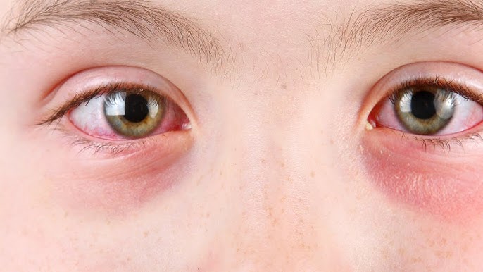
Eye Redness

Eye redness can often be a minor issue that is caused by the inflammation or irritation of blood vessels near the eye’s surface. This can be caused by an irritant, lack of sleep, or allergies. If the redness in your eyes is linked to some type of traumatic injury, you should see a doctor. Typical treatments for this condition are eye drops to help lubricate the eye, resting your eyes, or taking medication to treat your allergies. Eye redness can also be a warning sign for conjunctivitis, pink eye, or sun damage. These conditions should receive medical treatment from a licensed professional.
 

Night Blindness

Some people find that they have difficulty navigating in darker areas like movie theaters, dark rooms, or driving at night. Several possibilities could lead to night blindness. Some may have been born with this condition, or it could be the result of a degenerative retina. These issues cannot be resolved by a medical professional. However, night blindness can also be caused by nearsightedness, vitamin A deficiency, cataracts, or keratoconus. All of these issues can be corrected by a doctor.
 

Strabismus (Crossed Eyes)

Crossed eyes are not a condition that you can fix by yourself. This issue is seen in the eyes not properly lining up with each other. However, an optometrist may be able to help correct this issue, so it’s worth scheduling an appointment.
 

Nystagmus (Shaky Eyes)

Nystagmus is the constant shaking of the eyes of which the patient has no ability to control it. Medical professionals have several options for treatment at their disposal. Sometimes vision therapy is enough to strengthen the muscles and correct the issue. If this doesn’t work, surgery can also correct the problem. Your doctor can discuss your best options with you.
 

Colorblindness

Red and green color blindness is the most common form of colorblindness, although other types may affect your ability to see different color combinations. Color blindness is caused because the eyes lack the appropriate “cones” that help to interpret and differentiate these colors. This makes certain colors appear identical to colorblind individuals. Very severe forms of colorblindness only allow individuals to see shades of gray, but this is very rare.

While there is no current treatment for the correction of color blindness, special contact lenses or eyeglasses can be prescribed to help.
 

Uveitis

This condition is the inflammation of the uvea. There are several potential causes of uveitis that can be very serious, but uveitis is an umbrella phrase that covers all causes that create inflammation of the middle layer of the eye.

Some of these causes can be compromises of the immune system, like AIDS, rheumatoid arthritis, or ulcerative colitis. If you have light sensitivity, blurred vision, eye pain, and eye redness that lasts more than a few days you should see your eye care professional.
 

Knowing When To Get Help

Your eyes are a sensitive and critical organ for your health and small injuries or damage to your eyes can have lasting effects. For this reason, if you sense that something is wrong with your eyes, it’s always a good idea to visit your eye care professional. Additionally, make sure to get your regular annual eye exams which can help detect any issues that your eyes may be having and assure that you get the appropriate care more quickly.

Read More

Cataracts

A cataract can occur in either or both eyes, and most people will experience cataracts when they reach an older age. However, people can have an age-related cataract in their 40’s and 50’s, but during these years, most cataracts are small and do not affect vision. It is after age 60 that most cataracts begin to rob your vision.

The most common symptoms of a cataract are:

  • Cloudy or blurry vision
  • Colors seem faded
  • Glare. Headlights, lamps, or sunlight may appear too bright. A halo may appear around lights.
  • Poor night vision
  • Double vision or multiple images in one eye

Through a comprehensive eye exam, your Greenville EyeCare doctor can detect a cataract. If a cataract is detected, your doctor will recommend the best course of treatment. This may include treating an early cataract with new eyeglasses, recommending brighter lighting, use of anti-glare sunglasses, or even magnifying lenses. If surgery is required to remove the cloudy lens and replace it with an artificial lens, your doctor will review this treatment plan so that you are fully informed of the process and expectations.

Read More

Low Vision

Low vision is a term for conditions that result in reduced sight and cannot entirely be corrected with eyeglasses, contact lenses, medicines or surgery. Several eye diseases or conditions can cause low vision and here we will discuss the four most common causes of low vision and their risk factors.
 

Macular Degeneration

Macular degeneration is a disorder that affects the retina, which is the light-sensitive tissue lining the inside of the eye. Within the retina, the area responsible for sharp central vision (called the macula) deteriorates, causing blurred vision. This can cause a blind spot in the central area of vision, which leads to low vision.

There are two types of macular degeneration – non-exudative (dry form) and exudative (wet form). The dry form usually progresses slowly, while the wet form causes more rapid and severe vision loss due to abnormal blood vessels developing under the macula and leak fluid and blood. The biggest risk factor for macular degeneration is age. Other risk factors include genetics, race, smoking, and high blood pressure.
 

Diabetic Retinopathy

Diabetic retinopathy is a diabetes complication that affects the eyes. High blood sugar levels cause damage to blood vessels in the retina. These blood vessels can swell and leak (called macular edema). Sometimes they close, stopping blood from passing through (called macular ischemia). At first, diabetic retinopathy may cause no symptoms or only mild vision problems. However, over time, it can severely damage the retina, leading to low vision. Anyone who has diabetes can develop diabetic retinopathy. The risk increases for those who have had diabetes longer, as well as those with poor control of blood sugar levels, high blood pressure or cholesterol as well as those who smoke.
 

Cataracts

A cataract is a clouding of the lens in the eye that affects vision. This clouding can block the light from reaching the retina at the back of the eye, resulting in a general loss of vision. In some cases, a cataract can be surgically removed. Cataract surgery has a high success rate in otherwise healthy eyes but it is not always possible for people who also have other eye diseases. While the risk of cataract increases as you get older, other risk factors include diabetes, smoking, alcohol use, and prolonged exposure to ultraviolet sunlight.
 

Glaucoma

Glaucoma is a disease that damages your eye’s optic nerve. Most commonly, this occurs when fluid builds up in the front part of your eye which increases the pressure in your eye, damaging the optic nerve. There are four types of glaucoma: open-angle, normal tension, angle-closure, and secondary.
 

There are many risk factors for glaucoma, but those over 40 who have a family history of glaucoma should always have regular eye exams to check for glaucoma. In addition, you could be at an increased risk if your eye doctor has noted that you have high eye pressure, are farsighted or nearsighted, have thin corneas, thinning of the optic nerve, or have had an eye injury. Finally, certain health problems can cause glaucoma, such as diabetes, migraines, high blood pressure, or poor blood circulation.
 

How To Prevent Low Vision From These Diseases

Even though age and genetics play a part, you may have noticed some common risk factors – diabetes, smoking, and high blood pressure, which are all within your control.

Here are the most important steps you can take to reduce your risk of low vision from macular degeneration, diabetic retinopathy, cataracts, and glaucoma.

  • Be aware of your family’s health history
  • Get regular physicals
  • Move your body
  • Eat a healthy diet
  • Don’t smoke
  • Check out changes in vision with your eye doctor
  • Protect your eyes from the sun
  • Get your eyes checked at least every two years

By understanding the diseases that cause low vision, you can take steps now to reduce modifiable risk factors or get an early diagnosis to preserve your eyesight.

Read More

Thunderstorms & Seeing Rainbows

Summer, the season when everyone gloriously basks in the sun, soaks up that unparalleled pond life, and runs for cover when those tempestuous summer thunderstorms come barreling through. It’s also a time of rainbows!

Whether or not Pride Month was deliberately meant to be celebrated at the beginning of the rainbow season, that is serendipitously the way it turned out. Those summer sunny rainy days mean rainbows flapping in the summer breeze and arcing across the sky in all their ephemeral brilliance. But what if the rainbows you’re seeing aren’t on flags or fleeting on the horizon, but right there at the center of your vision, and not just in the heart of summer?

The quotidian flag and meteorological rainbows are entirely harmless and often extraordinary, but rainbows in your vision on a daily can be a sign of a serious eye condition.
 

Rainbow Vision

A lot of people will experience brief rainbow vision when exposed to bright lights, but the kind of rainbow vision that doesn’t dissipate relatively quickly or is accompanied by headaches, eye pain, blurred vision, or nausea is generally not something that should be dismissed.

The most common causes of rainbow vision are cataracts, glaucoma, migraines, and corneal edema.

At the top of the list are cataracts. Much like water droplets in the atmosphere create a haze through which sunlight is refracted creating a rainbow across the sky, a cataract is a clouding of the lens of the eye causing light to scatter creating rainbow-like halos around objects in your vision.

Glaucoma is likewise a condition of damage to the optic nerve caused by a variety of possible conditions, the result of which is a build-up of pressure and likely fluid inside the eye, distorting the eye, and creating rainbows around objects amongst other possible symptoms.

Migraines and corneal edema are both linked to increased pressure in the eye resulting in distorted vision including rainbows.
 

Treatment Of Rainbow Vision

There is no treatment for seeing rainbows after a sun shower or playfully fluttering on flag poles, but if the cause of your rainbow vision is something physiological, your eye doctor may recommend a few different avenues of treatment.

If cataracts are deemed the cause of your symptoms, an ophthalmologist may recommend cataract surgery to remove the cloudy lens and replace it with an artificial one. Beyond surgery, there are eye drops or other medications that can reduce inflammation or pressure in the eye clearing up symptoms like rainbows and headaches, or laser surgery may be recommended to treat glaucoma or other eye conditions.

Ultimately, if the rainbows you’re seeing are radiating around most of the objects in your vision, it’s time to contact your eye doctor for an exam, otherwise, enjoy the transient summer days and occasional rainbows until it’s time to hibernate again for winter.

Read More

Understanding Retinopathy

Retinopathy simply refers to damage to the retina caused by disease. The retina is the layer of tissue and cells at the back of the eye that captures light and transmits that light as images to the brain. Any damage to the retina via injury or disease can and often leads to vision impairment or complete vision loss.

Disease that affects the retina usually impacts the blood vessels in the retina, which results in loss of vision. The most common diseases to cause retinopathy, are hypertension and diabetes.

High Blood Pressure Damages The Retina

Healthy blood pressure is generally considered less than 120 systolic and less than 80 diastolic. The systolic number refers to the pressure when the heart contracts and pushes blood out through the body. This number is measured when the heart beats. The diastolic number is the opposite pressure when the heart relaxes and fills with blood returning from the body, which is measured between heartbeats. 

When the systolic pressure starts to push over 130 and that diastolic pressure over 80, a person starts to experience hypertension.

Hypertension or high blood pressure is usually caused by diets heavy in salts and fats, being overweight, not enough exercise and physical activity, over consumption of tobacco and alcohol, stress, and lack of sleep. High blood pressure can damage blood vessels throughout the body, including the retina where it is called hypertensive retinopathy, damage to the blood vessels in the retina resulting in vision impairment or loss of vision entirely.

Diabetic Retinopathy

The other most common cause of retinopathy is diabetes. Both type 1 and type 2 diabetes can ultimately cause diabetic retinopathy.

Diabetes often results in hyperglycemia, or high blood sugar, which if left untreated or unmanaged can lead to blockages or bulges in the blood vessels in the eye affecting essential blood supply and causing significant vision issues.

Retinopathy Prevention And Treatment

The best way to prevent retinopathy is to address the underlying diseases that cause damage to the retina. Managing and treating both hypertension and diabetes will help prevent damage to the retina and resulting retinopathy.

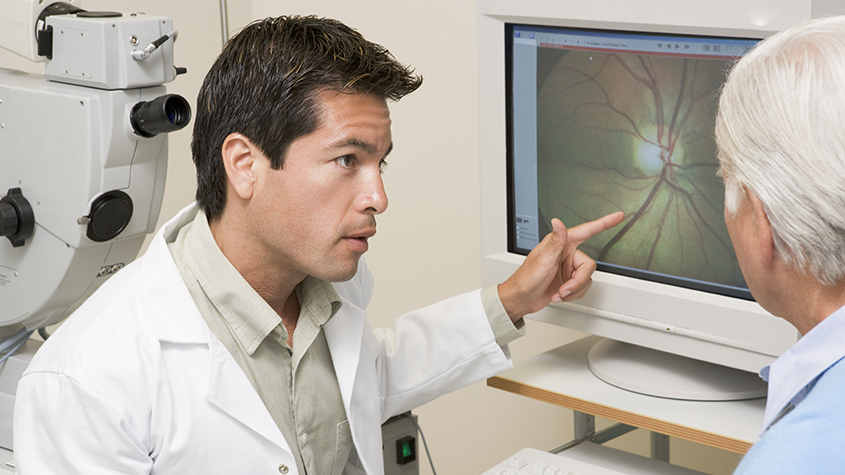
The next best way to prevent further damage and treat retinopathy is early diagnosis. An ophthalmologist will use both an ophthalmoscope during an exam and pre-screen with a Confocal Fundus Imaging System like the DRS Plus to closely examine the retina and observe for damage.

Blurry vision, double vision, floaters or spots in vision, unresolved eye pain, and decreased peripheral vision can all be early signs of retinopathy, though many patients do not experience significant symptoms before the damage is well established.

Regular comprehensive eye exams are always the most reliable way to prevent and treat any eye issues including retinopathy.

Read More

Glaucoma Treatment

If you’ve been diagnosed with glaucoma, you’re probably already familiar with the typical options in glaucoma treatment – eye drops, laser treatment or traditional surgery. While these are certainly effective, especially when glaucoma is diagnosed early, researchers have been working hard to offer new glaucoma treatments. Their goal is not only to improve outcomes but also reduce the treatment’s side effects and frequency of use.
 

What Is The Goal Of Glaucoma Treatment?

Before we dive into the new options, it’s important to understand the goal of any glaucoma treatment. At present, glaucoma is not curable. However, treatment can significantly slow the progression of the disease. Glaucoma damages your eye’s optic nerve. Extra fluid builds up in the front part of your eye (cornea), which increases the pressure in your eye. Reducing this pressure is the primary objective of any glaucoma treatment. This is often referred to as intraocular pressure or IOP.
 

What Are The Limitations Of Standard Glaucoma Treatments?

Eye drops for glaucoma treatment seem like an easy option but there are several challenges that can reduce its effectiveness. It can be difficult to get all the medicine in the eye, especially for older adults with less of a steady hand. In addition, since it must be applied daily, individuals may forget. Since the drops have no perceivable benefit because early stages of glaucoma have no symptoms, patients might make it a lower priority which is understandable since it may also have unpleasant side effects like burning, red eyes.

Beyond eye drops, laser surgery is a less invasive option. The laser opens clogged tubes and drains fluid. It can take a few weeks to see the full results. If laser surgery or drugs don’t relieve your eye pressure, you may need a more traditional operation. You would have to go into the hospital and will need a few weeks to heal and recover. Although usually effective, glaucoma surgery can make you more likely to get cataracts later on. It can also cause eye pain or redness, infection, inflammation, or bleeding in your eye.

What Are Recent Advances In Glaucoma Treatment?

Alternatives or Improvements to Eye Drops
The Glaucoma Research Foundation reported several new developments on the horizon. These technologies focus on reducing patient error in applying eye drops which would make the medication more effective and improve the quality of life for the patient. Here are some of the products underway:

  • A polymer, like a contact lens, would contain the drug; it would sit under the eyelid and release the medication over several months
  • Microneedles would inject medication into a specific spot to be most effective
  • Implantable extended-release devices using engineered highly precise microparticles and nanoparticles
  • Polymer-based intraocular delivery technologies that would allow customizable sustained release
  • Drops that allow the medication to get into the eye more easily
  • Tear duct plugs that release medication

In addition, people with glaucoma who take more than one eye drop per day are beginning to see those medications available as a single, combined eye drop. New products include Cosopt (timolol and dorzolamide), Combigan (timolol and brimonidine) and Simbrinza (brinzolamide and brimonidine).
 

Minimally Invasive Glaucoma Surgery

Minimally Invasive Glaucoma Surgery (MIGS) procedures are small cuts or micro-incisions through the cornea that cause the least amount of trauma to the surrounding tissues. Doctors implant a tiny device to allow fluid to drain from the eye, reducing internal pressure. Some devices (iStent) are implanted during cataract surgery. Cataract surgery alone lowers pressure, but the combination of both is more effective and can lower the need for medication.

These new techniques minimize tissue scarring, allowing for the possibility of traditional glaucoma surgery in the future if needed. They also give doctors the opportunity to treat patients earlier and more safely than older surgeries.
 

Monitoring Eye Pressure

An easy, accurate way to measure eye pressure is critical to monitoring the progress of glaucoma and adjusting treatment as needed. For patients that require more frequent testing of their eye pressure, there’s now an at-home tonometer called iCare HOME. There’s no puff of air and no eye drops. The patient can easily share the information with their eye doctor.

If you have a glaucoma diagnosis, you can feel confident that your glaucoma treatment options are only going to improve in the years ahead. Although the disease is not curable, it is very manageable with the right treatment.

Read More
TOP