Optometrists, Ophthalmologists, Opticians

Pediatric Eye Exams

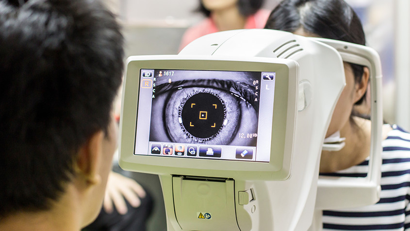
Regular eye exams are important for children since their eyes can change significantly in as little as a year as the muscles and tissue develop. Good eyesight is critical for a child’s life and achievements since success in school is closely tied to eye health. School demands intense visual involvement, including reading, writing, using computers, and blackboard/smartboard work. Even physical activities and sports require strong vision. If their eyes aren’t up to the task, a child may feel tired, have trouble concentrating, have problems in school or have difficulty playing their favorite games which may affect their overall quality of life.
 

When To Perform A Pediatric Eye Exam?

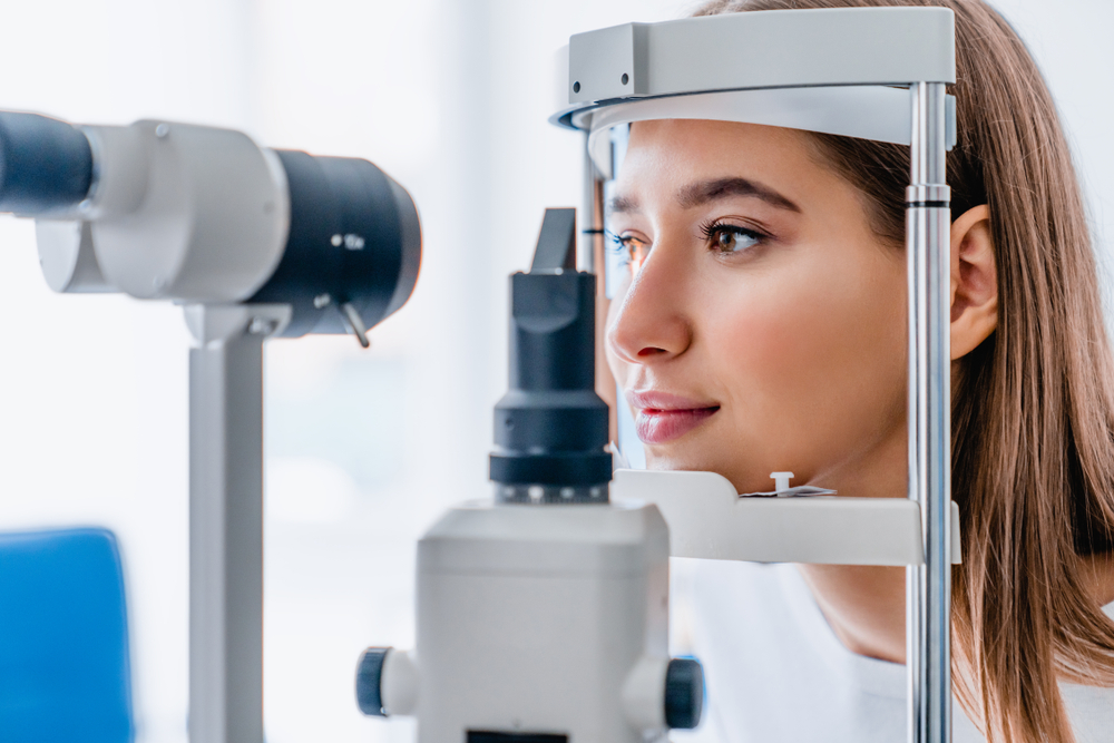
Routine eye and vision screenings should be performed on all children in order to help detect any abnormalities as their eyes develop. Then once they turn 18, they should have an exam every year.

For a newborn, an optometrist should examine the baby’s eyes and perform a test called “red reflex test” which is a basic indicator that the eyes are normal. In a case that the baby is premature or at high risk for medical problems for other reasons, has signs of abnormalities, or has a family history of serious vision disorders in childhood, the optometrist should perform a comprehensive exam.

A second eye health examination should be done to infants between six months and the first birthday. This examination includes tests of pupil responses to evaluate whether the pupil opens and closes properly in the presence or absence of light, a fixate and follow test to determine whether the baby can fixate on an object such as a light and follow it as it moves, and a preferential looking test which uses cards that are blank on one side with stripes on the other side to attract the gaze of an infant to the stripes and thus vision capabilities can be assessed. Infants should be able to perform this task well by the time they are 3 months old.

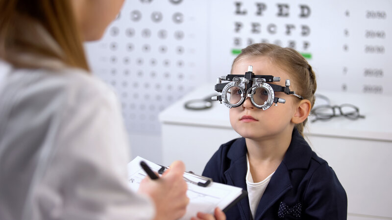
For a Preschooler, between the ages of 3 and 3½, a child’s visual acuity and eye alignment should be assessed. If the child is diagnosed with misaligned eyes (strabismus), “lazy eye” (amblyopia), refractive errors (astigmatism, myopia, hyperopia) or any other focusing problems, it’s important to begin treatment as soon as possible to ensure successful vision correction and life-long benefits.

At School age or upon entering school, the child’s eyes should be screened for visual acuity and alignment. In this age group, nearsightedness (myopia) is the most common refractive error and can be corrected with eyeglasses.
 

There are some signs that parents can tell if their child has a vision problem. For example, the child may squint, hold reading materials very close to their face, or complain about things appearing blurry. However, there are some less obvious signs that may indicate vision problems, such as having a short attention span, quickly losing interest in games, projects or activities that require using their eyes for an extended period of time, or losing their place when reading. As well as choosing to avoid reading, drawing, playing games or doing other projects that require focusing up close. Another sign is that a child may turn his or her head to the side when looking at something in front of them. This may be a sign of a refractive error, including astigmatism, so by turning their head helps the child see better.

That’s why it is so important for kids to have regular eye screenings with an optometrist. The earlier a vision problem is found and treated, the better off your child will be in and out of school.

Read More

Glaucoma Testing

You might be surprised at how many tests eye doctors use to diagnose glaucoma. A proper diagnosis requires careful evaluation of many aspects of your eye’s health – from eye pressure to cornea thickness to the health of your optic nerve. This article describes how your eye doctor assesses your risk and all the tests needed to properly diagnose glaucoma.
 

Risk Factor Assessment

Your eye doctor will begin by assessing your risk level for developing glaucoma. This will help determine the frequency and extent of testing needed. Through a family history and medical questionnaire, the eye doctor is looking for the following risk factors:

  • Over the age of 60
  • Ethnic background such as African or black Caribbean descent, Hispanic, or Asian
  • Family history of glaucoma, such as a sibling or parent with glaucoma
  • History of eye conditions, injuries or surgeries
  • Prolonged corticosteroid use (eye drops, pills, inhalers or creams)
  • Chronic conditions that affect blood flow, such as migraines, diabetes, low blood
    pressure or hypertension
  • Current or former smoker
     

If you’ve already had a comprehensive eye exam, your eye doctor will also consider these risk factors:

  • Eye pressure higher than normal (above 21 mm Hg)
  • Thin corneas (less than 0.5 millimeters)
     

Your type of eyesight is also important. People with farsightedness are at a higher risk for narrow-angle glaucoma, a more serious type that can advance quickly. While nearsightedness is associated with open-angle glaucoma, which progresses slowly without any symptoms.
 

Standard Glaucoma Tests

During a comprehensive eye exam, your eye doctor will always check for glaucoma, regardless of the risk level. This provides a baseline for comparison as you age. There are two tests: tonometry and ophthalmoscopy.
 

Tonometry

Tonometry measures the pressure within your eye. Your eye doctor will give you drops to numb your eyes. Then he/she will use a device called a tonometer, which applies a small amount of pressure with a warm puff of air.

Eye pressure is unique to each person, so it’s not always a reliable indicator for glaucoma. It’s simply another piece of information to help your eye doctor assess your eyes. The range for normal pressure is 12-22 mm Hg (“mm Hg” in millimeters of mercury, a scale for recording eye pressure). Most glaucoma cases are diagnosed with pressure over 20mm Hg. However, some people can have glaucoma at pressures between 12 -22mm Hg.
 

Ophthalmoscopy

This is an examination of your optic nerve. Your eye doctor will use eye drops to dilate the pupil, which makes it possible to see through your eye to examine the shape and color of the optic nerve. Then, using a small device with a light on the end, your optic nerve is magnified. Based on the results of these tests, your doctor may ask you to have more glaucoma exams.
 

Supplemental Glaucoma Tests

Perimetry

Perimetry is a visual field test. It creates a map of your complete field of vision. During this test, you’ll look straight ahead and then indicate when a moving light passes your peripheral (or side) vision. Try to relax and respond as accurately as possible. To ensure accuracy, your doctor may repeat the test to see if the results are the same the next time. If you’ve been diagnosed with glaucoma, a visual field test is usually recommended at least once per year to assess changes to your vision.
 

Gonioscopy

This diagnostic exam helps determine the angle of your iris and cornea. First, you’ll receive eye drops to numb the eye. A hand-held contact lens is gently placed on the eye. A mirror on the contact lens shows the doctor if the angle is closed and blocked (a possible sign of angle-closure or acute glaucoma) or wide and open (a possible sign of open-angle, chronic glaucoma).
 

Pachymetry

Last, your eye doctor may want to use pachymetry as another way to confirm a diagnosis. Pachymetry measures the thickness of your cornea, the clear window at the front of the eye. A probe called a pachymeter is gently placed on your cornea to measure its thickness. Pachymetry can help clarify your diagnosis because corneal thickness has the potential to influence eye pressure readings.

Glaucoma diagnosis is not as simple as you might expect. Be sure to have regular eye exams, especially if you have any of the risk factors, to detect glaucoma early.

Read More

Visual Field

Visual field testing is an important part of most standard comprehensive eye exams. Also sometimes known as perimetry testing, Visual field testing is a method to measure the entire scope of vision of an individual, including their peripheral/side vision.
 

The Importance Of Visual Field Testing

Visual field testing is one of the most effective diagnostic treatments in the detection of glaucoma. This is because when patients are affected by glaucoma, it is usually the peripheral vision that is affected by their condition first. However, it can also be used to detect central or peripheral retinal diseases, eyelid conditions such as drooping, optic nerve damage and conditions that affect the visual pathways from the optic nerve to the area of the brain where this information is processed into vision.
 

Visual field testing is also an important part of monitoring for people who are considered to be at risk for vision loss from disease and other problems, including those who have been diagnosed with the following:

  • Multiple sclerosis
  • Hyperthyroidism
  • Pituitary gland disorders
  • Central nervous system problems (such as a tumor that may be pressing on the brain)
  • Stroke
  • Diabetes
  • High blood pressure
     

What To Expect From Visual Field Testing

There are a variety of methods that can be used to perform visual field testing, including:
 

Static automated perimetry. This is where a machine is used to quantify how well the patient is able to detect flashing lights of varying size and brightness in different areas of their visual field, while they concentrate on a central point. The patient responds by pushing a button when they see the light.

Kinetic perimetry. This involves points of light that are fixed in size and intensity and are presented along the patient’s peripheral vision, before being gradually moved inwards to determine their field of vision. 

Visual field testing is non-invasive, painless and doesn’t require patients to have their eyes dilated. The results, which are usually presented in a series of charts, are digital and sent directly to your eye doctor for interpretation. Depending on the outcome of your results, you may be recommended for further diagnostic testing which could include blood tests. If you have been diagnosed with glaucoma, you will probably be recommended to have several visual field tests each year, which will help your eye doctor to monitor the progression of your condition and recommend treatments to slow it.

If you would like more information about visual field testing, or if you have concerns about your peripheral vision, please don’t hesitate to schedule an appointment with your experienced and knowledgeable eyecare team today.

Read More

Eye Emergencies

Eye emergencies cover a range of incidents and conditions such as; trauma, cuts, scratches, foreign objects in the eye, burns, chemical exposure, photic retinopathy, and blunt injuries to the eye or eyelid. Since the eye is easily damaged, serious complications can occur from an eye injury thus, any of these conditions without proper treatment can lead to a partial loss of vision or even permanent blindness. Likewise, certain eye infections, other medical conditions, such as blood clots or glaucoma, and eye problems such as a painful red eye or vision loss that are not due to injury also need urgent medical attention.
 

Depending On The Type Of Injury, Any Of The Following Symptoms May Be Present:

  • Bleeding or other discharge from or around the eye
  • Bruising
  • Decreased vision
  • Double vision
  • Loss of vision, total or partial, in one eye or both
  • Pupils of unequal size
  • Eye pain
  • New or severe headaches
  • Itchy eyes
  • Redness or bloodshot appearance
  • A sensation of something in the eye
  • Sensitivity to light
  • Stinging or burning in the eye
  • One eye is not moving like the other
  • One eye is sticking out or bulging
  • Nausea or headache occurring with eye pain (this may be a symptom of glaucoma or stroke).

A black eye is usually caused by direct trauma to the eye or face, causing a bruise resulting from bleeding under the skin. The skin around the eye turns black and blue, gradually becoming purple, green, and yellow over several days. Swelling of the eyelid and tissues around the eye may also occur. The abnormal color usually disappears within 2 weeks.

A blow to the eye can potentially damage the inside of the eye. Trauma is also a common cause of hyphemia, which is blood inside the front of the eye and is often due to a direct hit to the eye from a ball. Besides, certain types of skull fractures can cause bruising around the eyes, even without direct injury to the eye.

A chemical injury to the eye can be caused by a work-related accident, common household products such as cleaning solutions, garden chemicals, solvents, or other types of chemicals. Fumes and aerosols can also cause chemical burns. With acid burns, the haze on the cornea often clears and there is a good chance of recovery. However, alkaline substances such as lime, lye, drain cleaners, and sodium hydroxide found in refrigeration equipment may cause permanent damage to the cornea. It is important to flush out the eye with large amounts of clean water or salt water (saline).
 

Photic retinopathy, also known as foveomacular retinitis or solar retinopathy, is damage to the eye’s retina, particularly the macula, from prolonged exposure to solar radiation or other bright light, e.g., lasers or arc welders. It usually occurs due to staring at the sun, watching a solar eclipse, or viewing an ultraviolet, Illuminant D65, or other bright light. Immediate evaluation by your doctor is advised.

In case of an eye injury, cut or trauma, gently apply a clean cold compress to the eye to reduce swelling and help stop the bleeding. Do not, however, apply pressure to control bleeding. If blood is pooling in the eye, cover both eyes with a clean cloth or sterile dressing. And, call your doctor immediately.

In case of eye injury be sure NOT to:

  • rub or apply pressure to your eye
  • try to remove foreign objects that are stuck in any part of your eye
  • use tweezers or any other tools in your eye (cotton swabs can be used, but only on the eyelid)
  • put medications or ointments in the eye

As for contact lenses wearers, attempting to remove your contacts can make the injury worse. The only exceptions to this rule are in situations where there is a chemical injury and the lenses didn’t flush out with water, or where immediate medical help cannot be received.
 

How To Prevent Eye Injuries

Eye injuries can happen anywhere. Accidents can happen during high-risk activities, but also in places where you least expect them. There are things that can be done to decrease the risk of eye injuries, including wearing protective eyewear when using power tools or engaging in high-risk sporting events, following the directions carefully when working with chemicals or cleaning supplies, keeping scissors, knives, and other sharp instruments away from young children, and keeping a distance from amateur fireworks.

To decrease the chances of developing permanent eye damage, immediate medical evaluation is necessary in the event of an eye injury.

Read More
TOP