Optometrists, Ophthalmologists, Opticians

Optomap

Optomap is a new technology that gives eye doctors the ability to perform ultra-wide retinal imaging. In contrast to conventional retinal imaging, Optomap captures at least 50% more of the retina in a single capture, and with Optomap’s multi-capture function, up to 97% of the retina can be viewed. This gives eye care professionals greater opportunity to monitor the health and condition of patient vision.
 

Why Is Optomap Important?

Optomap is another great preventative eyecare technology tool. By allowing your eye doctor to have a comprehensive view of your retina, they will be able to detect any developing eye diseases early on, before they have a detrimental impact on your vision and day to day life. Not only can Optomap detect eye conditions such as retinal holes, retinal detachment, macular degeneration and diabetic retinopathy, but it can also be used to identify some general health conditions such as cardiovascular disease, stroke and cancer.
 

What To Expect From Optomap Scanning

Optomap is a fast, painless and non-invasive procedure that is suitable for patients of all ages, even children and pregnant women. Many patients require their eyes to be dilated ahead of the scan and will be given eyedrops which will widen their pupils and make it easier for the camera to see the structures inside the eye. Pupil dilation is painless, but patients may feel more sensitive to light both during their Optomap scan and afterwards for up to 24 hours. You may also have slightly blurred vision for a few hours. Once your eyes are dilated, you’ll be sat down and asked to look into a small device that will take the pictures of your retina. A short flash of light will let you know that the image has been taken, and the entire imaging is over in just a few seconds. The results will be sent digitally to your eye doctor who will then evaluate them. The results will also be stored on your personal optical record for future information.  

Read More

Routine Eye Exams

You may wonder, why do I need routine eye exams? Well they are an important aspect of maintaining one’s overall health. Regardless of how good your eyesight is, scheduling a regular eye exams is a great way to stay on top of your overall eye/vision health.

Adults should have an eye exam every year, depending on any existing vision problems, eye conditions or being diagnosed with significant risk factors, such as diabetes, high blood pressure, thyroid disease, previous eye injuries or family history. The doctor will recommend a frequency for routine follow-up exams based on the patient’s medical history. For instance, a diabetic patient will need a dilated eye exam every year while contact lens wearers need exams every year in order to look for changes that might affect lens fit and eye health.

Regular eye exams will also ensure that prescriptions for glasses or contact lenses are current as well as offer an opportunity to check for early signs of certain diseases. 

It may be important to see a doctor more frequently if one is experiencing any of the following:

  • Blurry vision or loss of vision.
  • Difficulty seeing things near and/or far away and perform basic tasks.
  • Flashing light in the eye.
  • Eye floaters, or small spots that appear in vision.

Why Should You Have Regular Eye Exams?

While eye exams are important for one’s vision, routine eye exams can also help to identify a variety of problems ranging from cognitive decline to diabetes. Since the eye is an extension of the brain and the only part of the body where blood vessels and tissue are visible, it allows an eye doctor to detect warning signs of the early stages of different health problems, such as diabetes which can present as bleeding in the eye or swelling in parts of the retina.

Besides diabetes, there are several other health problems that may be detected during a routine eye exam such as brain tumors that may cause swelling of the optic nerve and rheumatoid arthritis or other autoimmune disorders which may be the reason behind dry eyes.

Skin cancer on the eyelid is another health risk as the eyelid is very sensitive to ultraviolet rays and may be one of the first places affected by different types of skin cancers. Any spots or affected areas may be detected before skin cancer can spread to other parts of the body.

Addition, high blood pressure which would show as blood vessels in the back of the eye appearing bent or leaking, the narrowing of the vessels in the retina, swelling of the optic nerve, and hypertensive retinopathy in its earliest stages can be looked for during the exam.

There are also some progressive eye diseases that are not immediately apparent and should be tested for during regular eye examinations including:

  • Glaucoma which is the buildup of pressure within the eye that causes damage to the optic nerve and can lead to a loss of peripheral vision or a complete loss of vision. Glaucoma is a chronic, progressive eye disease that doesn’t show any symptoms or pain in the initial stages.
  • Macular degeneration is an eye condition that causes damage to the retina.
  • Cataracts which is the most common cause of blindness in the world. Cataracts occur when the lens of the eye becomes less flexible with age. Blurred or foggy vision and sensitivity to light are common symptoms. Cataracts are easily corrected with outpatient surgery.

Vision changes can have a profound effect on a person’s day-to-day life, but staying ahead of the curve can help ensure a lifetime of clear sight.

Read More

Pediatric Eye Exams

Regular eye exams are important for children since their eyes can change significantly in as little as a year as the muscles and tissue develop. Good eyesight is critical for a child’s life and achievements since success in school is closely tied to eye health. School demands intense visual involvement, including reading, writing, using computers, and blackboard/smartboard work. Even physical activities and sports require strong vision. If their eyes aren’t up to the task, a child may feel tired, have trouble concentrating, have problems in school or have difficulty playing their favorite games which may affect their overall quality of life.
 

When To Perform A Pediatric Eye Exam?

Routine eye and vision screenings should be performed on all children in order to help detect any abnormalities as their eyes develop. Then once they turn 18, they should have an exam every year.

For a newborn, an optometrist should examine the baby’s eyes and perform a test called “red reflex test” which is a basic indicator that the eyes are normal. In a case that the baby is premature or at high risk for medical problems for other reasons, has signs of abnormalities, or has a family history of serious vision disorders in childhood, the optometrist should perform a comprehensive exam.

A second eye health examination should be done to infants between six months and the first birthday. This examination includes tests of pupil responses to evaluate whether the pupil opens and closes properly in the presence or absence of light, a fixate and follow test to determine whether the baby can fixate on an object such as a light and follow it as it moves, and a preferential looking test which uses cards that are blank on one side with stripes on the other side to attract the gaze of an infant to the stripes and thus vision capabilities can be assessed. Infants should be able to perform this task well by the time they are 3 months old.

For a Preschooler, between the ages of 3 and 3½, a child’s visual acuity and eye alignment should be assessed. If the child is diagnosed with misaligned eyes (strabismus), “lazy eye” (amblyopia), refractive errors (astigmatism, myopia, hyperopia) or any other focusing problems, it’s important to begin treatment as soon as possible to ensure successful vision correction and life-long benefits.

At School age or upon entering school, the child’s eyes should be screened for visual acuity and alignment. In this age group, nearsightedness (myopia) is the most common refractive error and can be corrected with eyeglasses.
 

There are some signs that parents can tell if their child has a vision problem. For example, the child may squint, hold reading materials very close to their face, or complain about things appearing blurry. However, there are some less obvious signs that may indicate vision problems, such as having a short attention span, quickly losing interest in games, projects or activities that require using their eyes for an extended period of time, or losing their place when reading. As well as choosing to avoid reading, drawing, playing games or doing other projects that require focusing up close. Another sign is that a child may turn his or her head to the side when looking at something in front of them. This may be a sign of a refractive error, including astigmatism, so by turning their head helps the child see better.

That’s why it is so important for kids to have regular eye screenings with an optometrist. The earlier a vision problem is found and treated, the better off your child will be in and out of school.

Read More

Lens Technology

Thanks to the advancement of lens technology, glasses lenses are no longer a single, one size fits all solution. There are a variety of different lens types that can be used in glasses, giving patients greater flexibility and control over their vision than ever before.
 

Single Vision Lenses

Also known as monovision lenses, these lenses are designed to correct the wearer’s vision at just one distance, and have a single prescription covering the entire surface of the lens. They are most often recommended for people who are either nearsighted (myopia) or farsighted (hyperopia) and who need glasses for a specific activity, such as driving or reading.
 

Progressive Lenses

Progressive lenses are multifocal lenses that can correct a patient’s vision at different working distances, ranging from far distance to reading distance. However, rather than designating different areas on the lenses for different distances with visible lines separating them, progressive lenses have a gradual change so that the wearer can smoothly transition from one lens power to another.
 

Bifocal And Trifocal Lenses

As you may have guessed from the name, bifocal and trifocal lenses have either two or three lens powers depending on which type you choose. Bifocal lenses support distance vision in the top half of the lens, and near vision in the lower half. Trifocal lenses support distance vision in the top third of the lens, intermediate vision in the middle segment and near vision in the bottom third. Whichever variety you choose, you will see visible lines separating each segment.

Bifocal and trifocal lenses are recommended for patients who are near or farsighted, and those who develop presbyopia, which is the natural hardening of the eye lens, that occurs as we get older. Presbyopia makes it harder for the lens of the eye to adapt to focus at different distances.
 

Multifocal Lenses

Multifocal lenses are the alternative name given to bifocal, trifocal and progressive lenses.
 

Computer Lenses

Computer lenses are prescription lenses that are specifically designed to be worn when doing computer work. This is because they place the optimum lens power for viewing your computer screen exactly where you need it – which is closer than intermediate vision, but further away than reading material is usually held. Wearing computer lenses can significantly reduce the negative effects caused by the high visual demands of computer work, including blurred vision, redness, dry eyes, double vision and dizziness.
 

Transition Lenses

Also known as photochromic lenses, transition lenses are a special type of lens that darken when in the sunlight and lighten when in softer light or the dark. This versatility gives the wearer the convenience of being able to move between different environments without needing to change their glasses. This makes them extremely cost effective and prevent the wearer from needing to take multiple pairs of glasses out with them. Transition lenses also filter out many of the harmful UV rays that are emitted from the sun, helping to keep eyes healthy too. They are ideal for people who spend a lot of time going between inside and outside, or who work outside in varying weather conditions.
 

Blue Light Lenses

Blue light lenses are specially crafted lenses that contain filters that block out much of the artificial blue light that is produced by digital devices like computers, smartphones and tablets. Natural blue light is actually good for balancing our sleep-wake cycle, boosting our mood and enhancing our cognitive abilities so that we can function better day to day. However, too much blue light, especially from artificial sources, can have the opposite effect. Many people who fail to use blue light lenses can go on to develop digital eye strain, which produces symptoms like eye fatigue, dry eyes, blurred vision, headaches and more. Blue light lenses are recommended for anyone who spends a lot of time working on a digital device.
 

Polarized Lenses

Polarized lenses are used to reduce eyestrain and improve the quality of vision in patients on especially sunny days, making them ideal for anyone who spends a lot of time outdoors. They can do this because they have a special filter that blocks some of the light from passing through the lens. Vertical light is allowed to pass through, while horizontal light, such as that which bounces off of water and can be blinding, is blocked. Polarized lenses are most often used in sunglasses since they are worn outdoors, and the wearer also needs to protect their eyes from UV damage.

Read More

Face Types & Eye Glass Frames

When it’s time to select a new pair of eyeglasses, it can be challenging to find the frame that feels the best for you. Eyeglass makers have become increasingly aware of their clients’ desire to customize frames as well as offer a variety of colors, designs, and frame shapes. Understanding more about your face shape and the types of frames that would look best on you can help to reduce the amount of time you spend trying on frames that don’t complement your face shape. Here are a few useful tips to help you find glasses best suited for your face shape.
 

Face Types

While there are thousands of different frame shapes, there are only seven different face shapes.
 

Round

This face type is as wide is it is long. There are no specific or well-defined angles to this face type. Most people with this facial type look for frames that will elongate their face in order to draw out their natural features as well as give their face a thinner appearance. A good choice is often to select a frame that is angular and narrow.

Heart

If you can imagine the shape of a traditional heart, this face shape is easy to identify. The top 1/3 of the face is wider while the rest of the face tapers to a narrow chin. Individuals with this face shape will often seek frames that are wider at the bottom of the lens. This helps to balance the face and give the appearance of a narrower face at the top. Another great fit for this face type would be rimless frames.
 

Base Down Triangle

This face type contains a wide jaw and cheekbones but is narrow at the forehead. Cat-eye type frames are great for this face type, as are frames that are heavier near the eyebrow to provide more balance.
 

Diamond

This face type is the rarest. The diamond face is like the base-down triangle but differs because it has a narrow chin, thus appearing like a diamond. When selecting eyeglasses, look for oval-shaped frames, or frames that have more detail along the brow line.
 

Oblong

This face type looks like an oval, but these individuals have a straight cheek line. This face is fuller than it is long. Frame types that add some depth are ideal for this face. This can be done by finding frames that have decorative pieces along the temples or that have more depth with a reduced width.
 

Oval

Oval faces are the most common face type. They are considered to have balanced proportions so that most frame types will work well. An oval face is longer than it is wide and includes plump cheekbones. Walnut shaped eyeglasses are an ideal choice, but frames that are as wide or wider than the broadest part of your face would work as well.
 

Square

These individuals have a broad forehead and jaw that gives the appearance of the face having equal and straight sides. These face types usually do well with narrow or oval-shaped frames which will help to soften their features.
 

Additional Tips

Your hair color may play a role in the best frame color for you. Individuals with warm hair colors (brown or black hair) do best to choose a lighter colored frame to add contrast. People that have hair on the lighter end of the spectrum can pick just about any frame color that they would like and may look to other features like eye color or skin tone to help make their decision.

Skin tone has the opposite effect from hair color on the best choices for the frame color. Those with lighter skin hues do best with similar frame colors, and those with warmer skin tones can pick and choose options that they enjoy the most.

Above all else, you should pick a frame that you like best. There may be additional factors to consider when choosing the right frame for you, but perhaps the most important factor is your overall happiness and comfort.

Read More

Eye Safety Tips

The eye is a very delicate and fragile part of the body. According to one study on distance vision, about 188.5 million individuals are said to suffer from mild vision impairment, 217 million individuals suffer from moderate and severe impairment while 36 million individuals are blind. For near vision impairment, there are about 826 million individuals suffering. In all, there are about 1.3 billion people living with one of these impairments.

While a lot of people are aware that the eyes are an integral part of the body system, most of them do not pay very close attention to taking care of their eyes and are, in essence, taking it for granted. As the saying goes, “You don’t know what you have until it’s gone,” and therefore, we have decided to take the necessary steps to provide you with information on how to best to take care of your eyesight.
Below are ways to keep your eyes safe:

1. Eat Healthy
Eating a healthy diet is a very integral part of keeping the eyes healthy. Nutrients like lutein, vitamins E and C, omega-3 fatty acids, and zinc may help in preventing eye-related problems like cataracts and macular degeneration. These nutrients can be found in vegetables like collards, kale, spinach, fish like tuna, salmon, and halibut, as well as in beans, nuts, eggs, oranges, pork, and oysters.

Besides eating meals rich in nutrients, it is equally important to eat a well-balanced diet. Obesity can cause diabetes and other medical conditions, leading to blindness or glaucoma. Obesity is also regarded as the chief cause of blindness across the globe which should raise a red flag to watch your weight. You can always visit your doctor to get help with drafting a healthy eating plan.

2. Know your family’s medical history
Eye issues are usually hereditary which is why it is important to collect a family history on any diseases that may affect eye health and then report them to your eye doctor. This will help to determine your chances of any future eye conditions as well as come up with an action plan for future eye exams.

3. Avoid Smoking
Smoking, in general, is harmful to your overall health. In regards to your eyes, it can cause macular degeneration, destroy the optic nerve, and cause cataracts. It can even lead to vision loss. Quitting smoking if you already smoke, or better yet, not even starting, will be extremely beneficial to your overall health and wellness.

4. Wear Sunglasses
Sunglasses are more than just fashion accessories, they are vital in protecting the eyes from ultraviolet rays emitted from the sun. UV rays can cause macular degeneration and even cataracts which is why it is important to wear sunglasses that protect against 99% -100% of UV rays.

5. Obey workplace safety precautions
It is important we wear protective eye gears when engaging in sporting activities or jobs that could be harmful to the eyes. Such eyewear includes safety goggles, glasses, and helmets. Most of these protectors are from polycarbonates, which is a much stronger material than plastic.

6. Rest your eyes
Just like our bodies, the eyes also need rest to perform optimally. Depriving yourself of adequate sleep and rest can have an adverse effect on the eyes. During the use of the computer, remember to rest the eyes for at least 20 seconds after every 20 minutes by looking away from the screen. Doing this will reduce eye strain.

7. Visit your eye doctor regularly
Getting regular eye exams is one of the most important things you can do for your eye health. Conditions like glaucoma are much easier to treat if they are discovered early so don’t stay far from your doctor. 
 

First Aid

Eye injuries can happen at any time and it is good to know how to handle such emergencies until you can reach a medical professional.

  • If you get hit on the eyes, you should put a shield like a Styrofoam cup on the bone of the eyes. Avoid putting pressure on the eye
  • If something enters your eyes, avoid removing it as it may lead to the tearing of important tissue in the eyes. A Styrofoam cup would also do the job here until you can get to a medical professional.
  • In the case of a black eye, putting an ice pack on the eye will help to reduce bruising and swelling
  • If it’s a chemical burn, rinse the eyes with clean water for about 30 minutes, holding your head beneath the tap. While rinsing, keep your eyes wide open, rolling your eyeballs in all directions so as to ensure the water gets to every part.
     

So give some of these tips a try and remember to schedule regular eye exams. Prevention is always a better alternative to trying to find a cure.

Read More

Common Ocular Diseases

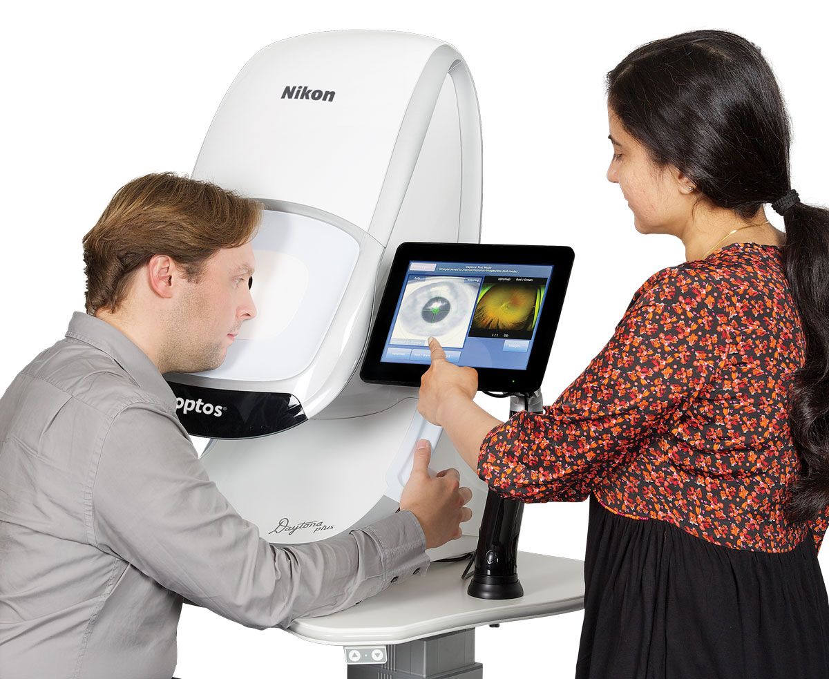
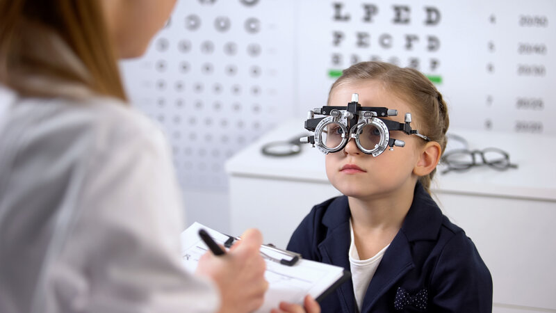
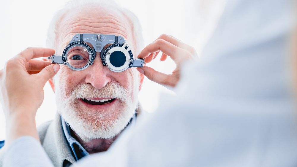
Both optometrists and ophthalmologists treat many common types of ocular disease. However, for the best outcome, it’s important to see an eye doctor regularly. They can identify any issues before they become serious problems.

Fortunately, they can treat all of the diseases mentioned below, and in some cases, you can do certain things to prevent them from developing. Look at the most recent statistics, and you’ll see why good eye health care matters.

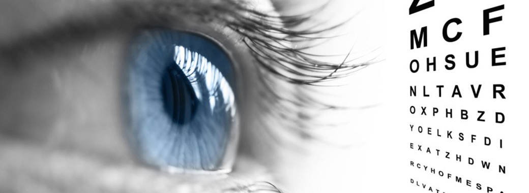
Currently, more than 4.2 million people in the U.S. alone over the age of 40 are partially blind or have poor visual acuity. Although a lot of things cause these problems, the ocular diseases listed below are the most common.

Macular Degeneration

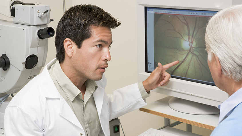
This is commonly referred to as “age-related macular degeneration” because it affects seniors. Not only does it cause blurriness and distortion but left untreated, individuals lose their central vision. In other words, they are unable to see anything through the center portion of the eye.

Two types of this ocular disease exist. First, wet macular degeneration means that abnormal blood vessels that are located behind the retina grow under the macular. Along with leaking blood and fluid, this leads to scarring and, sometimes, permanent damage. Second, dry macular degeneration progresses slowly as part of the natural aging process. Typically, it affects both eyes at some point.

Cataracts

Roughly 20 million people in the U.S. over the age of 40 have cataracts in either one or both eyes. While they can develop in children, teens, and young adults, cataracts are most often associated with age. With this, a film covers the eye, which, in turn, makes everything appear blurry.

Of all the different kinds of ocular diseases that lead to blindness worldwide, cataracts rank number two. Fortunately, an eye doctor can remove the damaged lens, followed by implanting an artificial one. After recovery, patients see amazingly well.

Diabetic Retinopathy

If you have diabetes, then you’re at risk of developing this ocular disease. This particular disease causes progressive damage to the retina’s blood vessels. The first stage consists of mild non-proliferative retinopathy and then moderate non-proliferative retinopathy, which blocks some of the vessels.

Then, it moves into stage three or severe non-proliferative retinopathy, which means more blood vessels become blocked. The fourth and final state, proliferative retinopathy, is the most advanced. Although Diabetic Retinopathy does affect just one eye on occasion, it typically involves both eyes.

Start by improving your overall health. Eat balanced meals, keep your blood pressure and cholesterol levels down, and take insulin. In addition, regular exercise, losing weight, and giving up smoking all make a huge difference. From there, a qualified eye doctor can provide you with treatment options to reduce the risk of losing your vision.

Glaucoma

Many people think glaucoma is one type of ocular disease. However, it’s a group of diseases that cause damage to the optic nerve. When that happens, people face the risk of losing their sight completely. With glaucoma, the fluid pressure inside the eyes gradually rises.

There are also two categories of glaucoma: open-angle and closed-angle. Not only is open-angle glaucoma chronic, but it also progresses slowly. Often, a person can have this type without knowing it. Unfortunately, they don’t realize there’s an issue until they have a comprehensive eye exam performed.

As for closed-angle glaucoma, it’s usually painful and it comes on suddenly. In addition, an individual can lose their vision much faster with this kind of glaucoma compared to the open-angle kind. Because this happens fast and involves pain, it’s diagnosed much quicker as well.

For these common types of ocular diseases, it’s important to have your vision checked. If an ocular disease is diagnosed, your optometrist will be able to determine the best treatment plan for optimal eye health and vision.

Read More

Diabetic Related Eye Exams

You have almost certainly heard of diabetes, which is one of the most common chronic health conditions in the United States with an estimated 100 million adults currently living with diabetes or pre-diabetes. This metabolic disorder occurs when the body is no longer able to regulate its own blood sugar levels and requires intervention to keep them stable. Most people are aware that diabetes can have serious consequences for our health. However, you may be surprised to learn that it can also influence our vision. This is because patients who are diabetic can go on to develop a complication that is known as diabetic retinopathy. Without prompt treatment, diabetic retinopathy can cause permanent vision loss. It is for this reason that patients who suffer from diabetes are asked to attend regular diabetic-related eye exams. 
 

What Is Diabetic Retinopathy?

For us to be able to see clearly, our eyes need to be healthy and functioning perfectly. The most important component of our eyes are the retina. Found at the very back of the eye, the retina is a patch of light-sensitive cells that have the job of converting the light that passes into the eye into messages that are passed up the optic nerve and into our brain. Our brain then receives them and tells us what we can see and how clearly we can see it. 

The retina relies on a continuous supply of blood, which is delivered using a network of tiny blood vessels. Over time, having continuously high blood vessels can damage these blood vessels causing a leak of blood and other fluids onto the retina. If this happens, scarring may occur which could compromise the quality of your vision. 
 

Am I At Risk Of Diabetic Retinopathy?

Technically, anyone who suffers from diabetes, whether it be Type 1 or Type 2, could be at risk of developing diabetic retinopathy. However, the condition is more likely in certain situations. These include if:

  • your blood sugar levels are uncontrolled or poorly controlled
  • you have a long history of diabetes
  • you have high blood pressure (hypertension)
  • you suffer from high cholesterol 
  • you are pregnant

Regular diabetic-related eye exams will enable your eye doctor to monitor your condition and ensure that any signs of diabetic retinopathy are detected and acted upon immediately. 
 

What To Expect From Diabetic-Related Eye Exams?

The process of a diabetic eye exam is very simple and straightforward. In fact, in most instances, it is included within the other elements of comprehensive eye exam and you may not even realize that you have had a specific test to check for diabetes-related complications. 

Diabetic eye screening is non-invasive. You will be given eyedrops which will blur your vision. These may sting a little when they are administered, but this will pass within just a few moments. Once your vision is blurred, you will be asked to rest your head onto a device and stare down a lens. This leads to a camera which will take images of the backs of your eyes so that your eye doctor can assess the structures, which include the retina, for any abnormalities. You will see a flash when each image is taken, but at no point should you be in any pain. 

In addition to the images of the back of your eye being taken, you will also be given a visual acuity test. This is where you will be asked to read letters off a chart a short distance away, as well as reading from a card held in front of you. 

The information that your eye doctor will obtain from your examination will be able to tell them if you are experiencing any of the signs of diabetic retinopathy. If so, they will discuss the best way to get your condition under control. This could involve a combination of elements, including controlling your diabetes more effectively, taking medications or more invasive treatment to preserve your vision. Your eye doctor will give you more specific information based on your individual circumstances. 

If you have further questions about diabetic-related eye exams, please contact a knowledgeable eye care team. 

Read More

Scleral Lenses

Contact lenses come in a variety of shapes and styles to suit the range of needs for individual patients. What this means is that there is almost certainly a type of contact lens that will both feel comfortable for you and improve your vision. 
 

Some types of contact lenses are known as speciality lenses. This is because they are designed specifically to overcome some eye issues which may prevent a patient from wearing the most common and generic styles of contact lenses. In fact, speciality contact lenses have transformed the vision options of many patients who would otherwise have only had the choice between wearing glasses or undergoing laser vision correction. There are numerous types of speciality contact lens, including scleral lenses.
 

What Are Scleral Lenses?

Scleral contact lenses get their name from the fact that, unlike regular contacts, they vault over the entire corneal surface and rest on the white part of the eye, which is called the sclera. This makes them larger than standard contacts, which in turn provides a variety of benefits. Their size makes them easier to handle and more stable when on the eye, which in turn provides the patient with sharper and more reliable vision. They are also less likely to become dislodged and come out. 

Scleral contact lenses are also gas permeable, which means that oxygen can pass right through them and reach the surface of the eyes. This is important for comfort, particularly for patients who experience dry eyes. The gap that is created between the back of the contact lens and the front surface of the eye can also trap tear film, acting as a fluid reservoir which will keep the eyes moist and healthy. 
 

Types Of Scleral Lenses

Within scleral lenses there are several different sizes to choose between. These are based on where the lenses meet the surface of the eye and are as follows:

Full scleral lenses: the largest type, they provide the greatest amount of clearance between the cornea and lens and rest on the outer sclera. 

Mini scleral lenses: the mid-size variety, make contact with the eye on the anterior sclera. 

Semi-scleral lenses: although larger than conventional lenses, they are not huge, and the edge of the contact lens rests on the junction between the cornea and the sclera. 

Your scleral lens provider will be able to make a recommendation as to the correct size for you, which will be based on your individual requirements. 
 

Who Are Scleral Lenses Recommended For?

Although technically anyone can wear and benefit from scleral lenses, they are a particularly good option for patients who have eye conditions or irregularities that for which regular contacts are not suitable. This includes patients with:

Dye eye syndrome. Contact lenses float on a fine layer of tear film, which keeps them comfortable and in position. Patients with dry eye syndrome either do not have enough natural tear film, or what they do have drains away too quickly. This can
make wearing regular contact lenses difficult and uncomfortable. However, the design of scleral lenses keeps moisture on the surface of the eyes, eliminating some of the effects of dry eyes. This makes scleral lenses a great choice for patients with dry eyes. 

Irregular corneas. The cornea is the clear dome that covers the front part of the eye and many people have a cornea that is a slightly, or significantly, irregular shape. This causes light to be refracted incorrectly when it enters our eyes, triggering vision problems. Issues arising from irregularly shaped corneas cannot be completely corrected using glasses or conventional contact lenses. Therefore, scleral lenses, which provide sharper and more accurate vision, are recommended. 

Read More

Glaucoma Testing

You might be surprised at how many tests eye doctors use to diagnose glaucoma. A proper diagnosis requires careful evaluation of many aspects of your eye’s health – from eye pressure to cornea thickness to the health of your optic nerve. This article describes how your eye doctor assesses your risk and all the tests needed to properly diagnose glaucoma.
 

Risk Factor Assessment

Your eye doctor will begin by assessing your risk level for developing glaucoma. This will help determine the frequency and extent of testing needed. Through a family history and medical questionnaire, the eye doctor is looking for the following risk factors:

  • Over the age of 60
  • Ethnic background such as African or black Caribbean descent, Hispanic, or Asian
  • Family history of glaucoma, such as a sibling or parent with glaucoma
  • History of eye conditions, injuries or surgeries
  • Prolonged corticosteroid use (eye drops, pills, inhalers or creams)
  • Chronic conditions that affect blood flow, such as migraines, diabetes, low blood
    pressure or hypertension
  • Current or former smoker
     

If you’ve already had a comprehensive eye exam, your eye doctor will also consider these risk factors:

  • Eye pressure higher than normal (above 21 mm Hg)
  • Thin corneas (less than 0.5 millimeters)
     

Your type of eyesight is also important. People with farsightedness are at a higher risk for narrow-angle glaucoma, a more serious type that can advance quickly. While nearsightedness is associated with open-angle glaucoma, which progresses slowly without any symptoms.
 

Standard Glaucoma Tests

During a comprehensive eye exam, your eye doctor will always check for glaucoma, regardless of the risk level. This provides a baseline for comparison as you age. There are two tests: tonometry and ophthalmoscopy.
 

Tonometry

Tonometry measures the pressure within your eye. Your eye doctor will give you drops to numb your eyes. Then he/she will use a device called a tonometer, which applies a small amount of pressure with a warm puff of air.

Eye pressure is unique to each person, so it’s not always a reliable indicator for glaucoma. It’s simply another piece of information to help your eye doctor assess your eyes. The range for normal pressure is 12-22 mm Hg (“mm Hg” in millimeters of mercury, a scale for recording eye pressure). Most glaucoma cases are diagnosed with pressure over 20mm Hg. However, some people can have glaucoma at pressures between 12 -22mm Hg.
 

Ophthalmoscopy

This is an examination of your optic nerve. Your eye doctor will use eye drops to dilate the pupil, which makes it possible to see through your eye to examine the shape and color of the optic nerve. Then, using a small device with a light on the end, your optic nerve is magnified. Based on the results of these tests, your doctor may ask you to have more glaucoma exams.
 

Supplemental Glaucoma Tests

Perimetry

Perimetry is a visual field test. It creates a map of your complete field of vision. During this test, you’ll look straight ahead and then indicate when a moving light passes your peripheral (or side) vision. Try to relax and respond as accurately as possible. To ensure accuracy, your doctor may repeat the test to see if the results are the same the next time. If you’ve been diagnosed with glaucoma, a visual field test is usually recommended at least once per year to assess changes to your vision.
 

Gonioscopy

This diagnostic exam helps determine the angle of your iris and cornea. First, you’ll receive eye drops to numb the eye. A hand-held contact lens is gently placed on the eye. A mirror on the contact lens shows the doctor if the angle is closed and blocked (a possible sign of angle-closure or acute glaucoma) or wide and open (a possible sign of open-angle, chronic glaucoma).
 

Pachymetry

Last, your eye doctor may want to use pachymetry as another way to confirm a diagnosis. Pachymetry measures the thickness of your cornea, the clear window at the front of the eye. A probe called a pachymeter is gently placed on your cornea to measure its thickness. Pachymetry can help clarify your diagnosis because corneal thickness has the potential to influence eye pressure readings.

Glaucoma diagnosis is not as simple as you might expect. Be sure to have regular eye exams, especially if you have any of the risk factors, to detect glaucoma early.

Read More
TOP